

Brain Ultimate Neuronavigation
See exactly where you are treating.
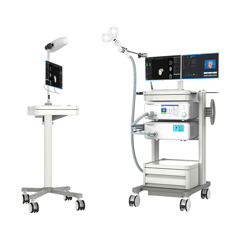
Brain Ultimate Neuronavigation tracks the treatment coil against a 3D model of your patient's head in real time — so every session lands on the same target, at the same angle, with confidence you can document.
- Real-Time Coil Tracking
- Personalized MNI Targeting
- Individual Imaging or Common Mode
- Session-to-Session Repeatability
At a glance
From Coordinate to Cortex
Optical tracking that connects your treatment plan to the patient in front of you.
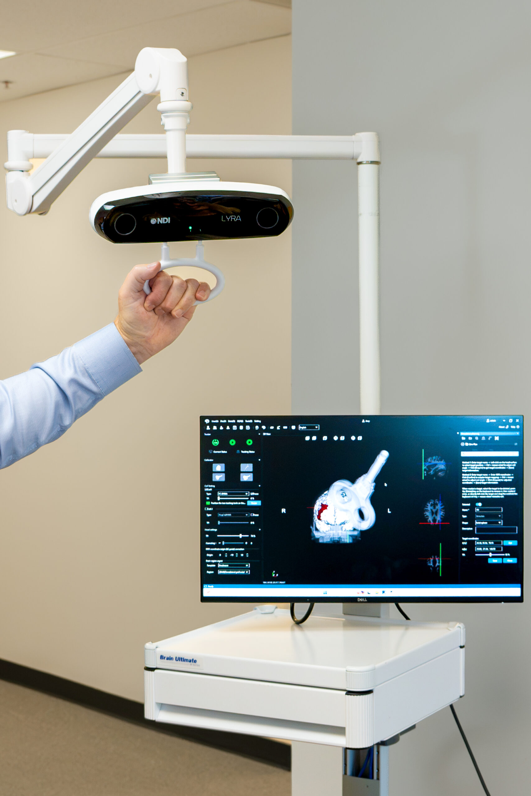
Neuronavigation uses an infrared optical camera to follow two tracking tools — one on the treatment coil, one on the patient's head — and registers both to a 3D head model. The result is a live picture of exactly where the coil sits relative to the intended target, updated continuously throughout the session.
-
Registered to the PatientA short calibration at the start of each session maps the patient's real-world anatomy to the on-screen model using standard landmarks.
-
Tracked ContinuouslyHead and coil positions are followed in real time. If either moves, the display moves with it.
-
Documented AutomaticallyEvery session writes to the treatment record, with reports generated from your chosen template.
The Right Spot at the Wrong Angle Is Not the Same Target
Location alone does not define a treatment site — and neither does the scalp.
A target is six values, not three
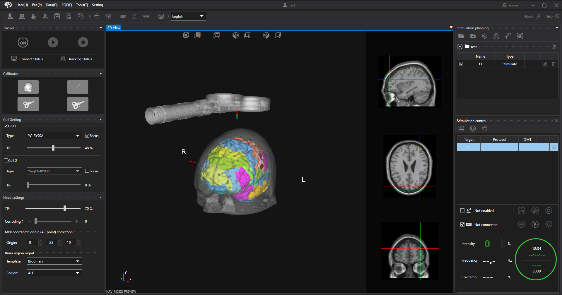
Position tells you where on the scalp the coil sits. Orientation — pitch, roll, and yaw — tells you how it is tilted and which way the handle points. Induced field strength and direction depend on both, so the same scalp location at a different coil angle delivers a different treatment. Neuronavigation tracks all six continuously, which is what makes a target reproducible from one session to the next.
And the scalp is not the cortex
Scalp-landmark methods estimate a cortical location from external measurements. They are quick, they need no imaging, and they are the reason TMS scaled. But head shape, cortical folding, and the depth from scalp to target all vary between people, so the same measurement lands in a different place on a different head.
What navigation adds
Navigation replaces an estimate with a measurement. The target is chosen once, in the patient's own anatomy, and then found again at every session rather than re-estimated at every session. A target worth choosing is only worth choosing if it can be found again at the next session — and finding it again is what a navigation system does.
Guidance That Holds Through the Session
Offset tracking keeps treatment inside the zone you define.
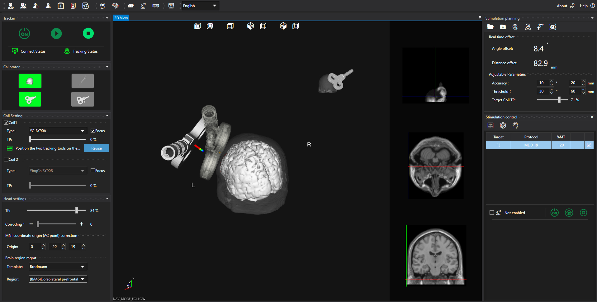
Once a target is set, the system displays live offset — how far the coil is from the target in distance, and how far off in angle. A semi-transparent guide coil shows the ideal position. Bring distance inside the accuracy zone first, then angle; small movements have a large effect once you are close.
Accuracy zone
The inner tolerance. Both distance and angle must be inside it before stimulation starts, and it is enforced for the whole session.
Threshold zone
The outer warning band. During an active session the coil may drift into it and stimulation continues, but repositioning is needed.
Set Targets the Way You Work
Standard sites, published coordinates, the patient's own imaging, or the position you just found by hand.
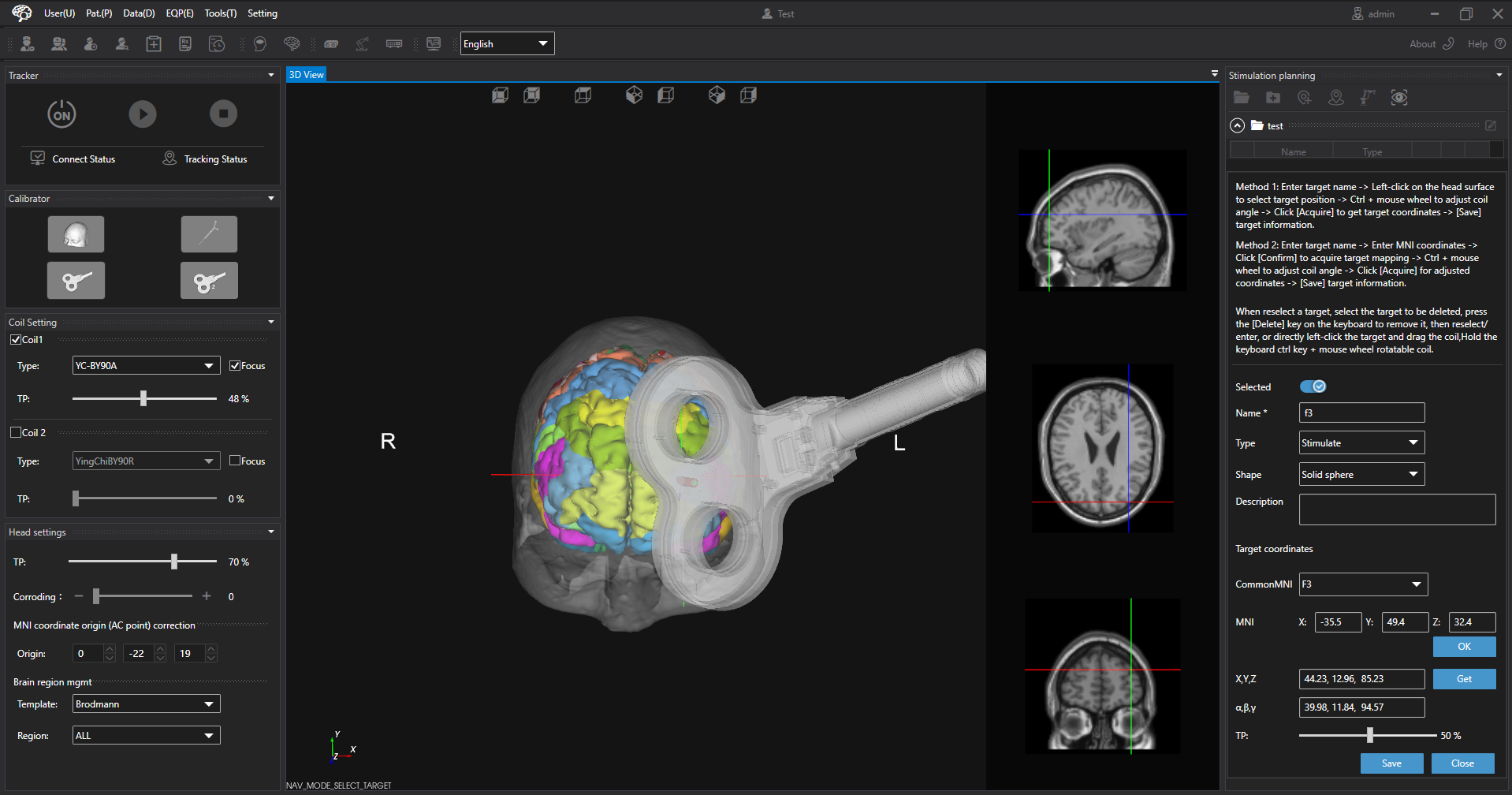
Standard 10-20 sites
Select a common site from the 10-20 dropdown, such as F3 for the left DLPFC.
MNI coordinates
Enter published coordinates directly, or scroll the slice views and click to capture a point.
The patient's own imaging
Import the patient's functional and structural scans and derive personalized MNI coordinates in the same software.
Directly on the model
Click the target on the 3D head, then drag and rotate the virtual coil to fine-tune position and angle.
Save current coil position
With calibration complete, hold the real coil where you want it and record that position as the target.
Imaging import — two formats, two behaviours
-
DICOMCarries both functional and structural data and produces a pre-made 3D model of the patient's head, ready to target against.
-
NIfTIImports as slices, navigated in the slice views.
-
Either format yields personalized MNI coordinatesComputed in the navigation software. No separate analysis package and no export step.
Head models
-
Common modeFor patients without imaging. Standard atlases: adult male, adult female, adolescent, and child.
-
Individual imagingThe patient's own scans, with anterior commissure alignment so MNI targets map to that patient's anatomy.
Navigation, Stimulation and Threshold in One Application
One application, one record, one workflow.
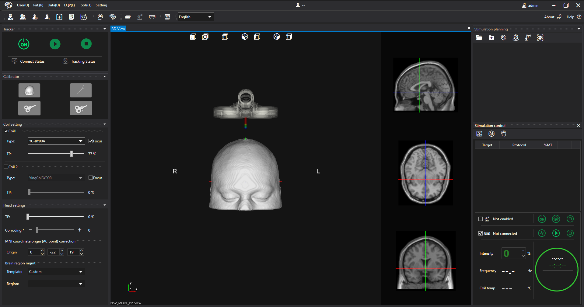
Brain Ultimate Neuronavigation is built by Brain Ultimate rather than integrated from a third-party navigation product. QuickVision handles all of it — navigation and targeting, stimulator control, protocol parameters, coil temperature, and motor threshold determination — in a single application. There is no second application to open, no window to switch between mid-session, and no separate record to reconcile afterwards.
In practice that means the clinician who is positioning the coil is looking at the same screen that is running the protocol. Target, offset, stimulator status and thermal state are read together rather than assembled from two systems that do not know about each other.
- Drives Brain Ultimate TMS protocols directly — rTMS, cTBS, iTBS, and paired-pulse
- Motor threshold with or without the MEP Module, including paired-pulse thresholds
- Live coil temperature and stimulator status alongside the target view
- Treatment records and reports generated in the same session
A Session, Start to Finish
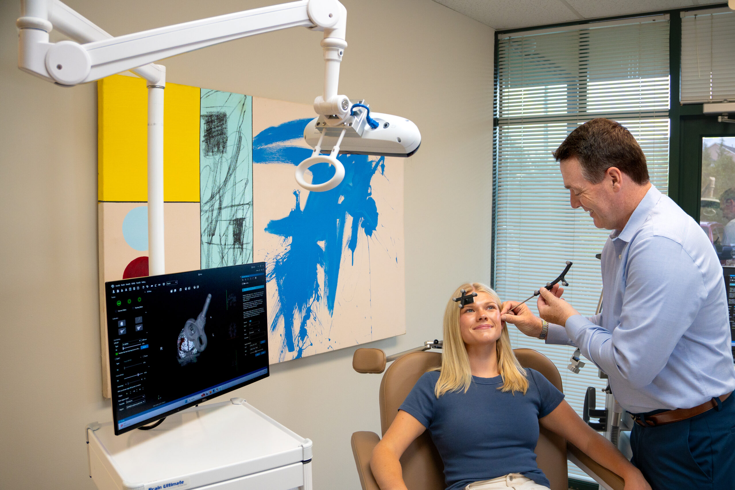
- Select the patientNew profile, or load an existing one with its saved plan and calibration.
- Load or create the planHead model, targets, and bound protocols in one container.
- Confirm motor thresholdDetermine it for new patients; returning patients carry a stored value.
- CalibrateHead first, then coil. Both indicators turn green before you continue.
- PositionBring distance, then angle, inside the accuracy zone and lock the arm.
- TreatStart, monitor, and stop. The session logs itself and the report is generated from the record.
Monitoring That Does Not Depend on Watching
Automatic Pause on Drift
Stimulation pauses if the coil leaves the threshold zone and resumes once it is back on target.
Thermal Protection
Coil and stimulator temperatures are monitored continuously, with automatic protective response at defined limits.
Calibration Integrity
The selected coil type must match the coil in use. A mismatch invalidates calibration, and the system is explicit about it.
What Ships With the System
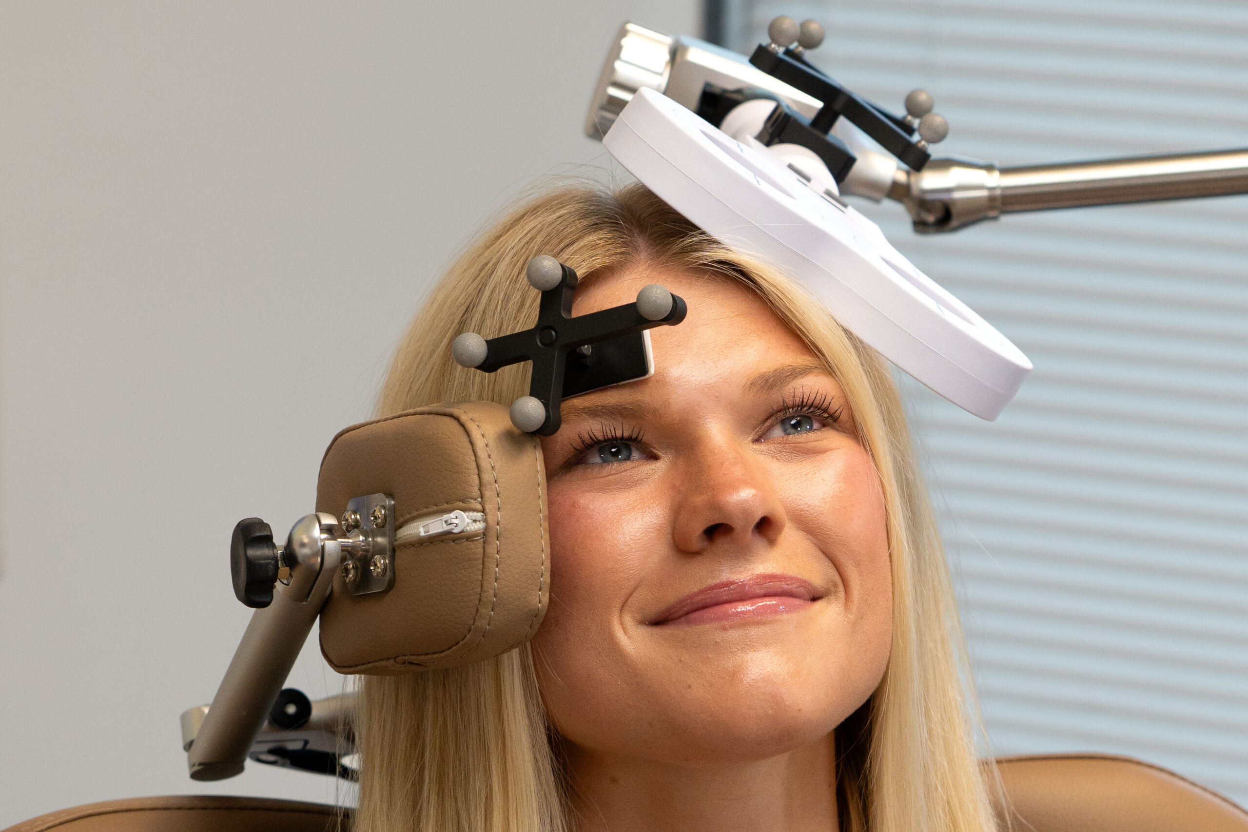
- Infrared tracking cameraFollows the coil and patient tracking tools throughout the session.
- Coil tracking toolMounts to the treatment coil with an adjustable bracket.
- Patient tracking toolHeadband or glasses, positioned opposite the treatment site.
- Calibration probeRegisters anatomical landmarks and coil reference points.
- Navigation workstationDual-display host running the QuickVision software, with dark and light themes and English language support.
System Specifications
| Item | Detail |
|---|---|
| System type | 3D optical neuronavigation for transcranial magnetic stimulation |
| Tracking | Infrared optical, coil and patient tracking tools |
| Coordinate space | MNI, with anterior commissure alignment on individual models |
| Head models | Common mode standard atlases (adult male, adult female, adolescent, child) or individual patient imaging |
| Imaging formats | DICOM — functional and structural, produces a pre-made 3D head model. NIfTI — imports as slices. |
| Personalized targeting | Personalized MNI coordinates derived from the patient's own imaging, computed in the navigation software |
| Software | QuickVision — navigation, targeting, stimulation control and motor threshold in a single application |
| Protocols | rTMS, cTBS, iTBS, and paired-pulse variants |
| Motor threshold | With or without MEP Module; single and paired-pulse; 2- or 4-channel acquisition |
| Pairs with | Brain Ultimate TMS (M-Series) and the MEP Module |
| Reporting | Treatment records with template-based report generation and batch export |
| Regulatory | FDA 510(k) cleared — K261228 |
| Pricing | Contact for pricing |
Regulatory Statement
Brain Ultimate Neuronavigation is FDA-cleared under K261228 for spatial positioning and orientation of the treatment coil for transcutaneous magnetic stimulation systems under the supervision of a tracking system.
Stimulation is delivered by the Brain Ultimate TMS system, which holds three separate 510(k) clearances: K230735 for Major Depressive Disorder and K243459 for Obsessive-Compulsive Disorder, which are its cleared indications, and K243460 for intermittent theta burst stimulation, which is a cleared modality.
Navigation supports the delivery of cleared protocols. It does not extend the cleared indication set.
Product documentation supplements, and never replaces, the Instructions for Use. Consult the IFU for warnings, contraindications, and full regulatory information.
Bring Precision Targeting to Your Practice
Neuronavigation is available for new Brain Ultimate TMS systems and as an addition to systems already in service. Contact us for pricing and configuration.