

Introducing Brain Ultimate Neuronavigation: Precision Targeting for TMS
We're excited to announce that Brain Ultimate Neuronavigation, our newest addition to the M-Series Ultimate platform, has received FDA clearance (K261228) for spatial positioning and orientation of the treatment coil during TMS. It's available now as an add-on for existing Brain Ultimate TMS systems or bundled with a new M-Series Ultimate purchase.
What Is Brain Ultimate Neuronavigation?
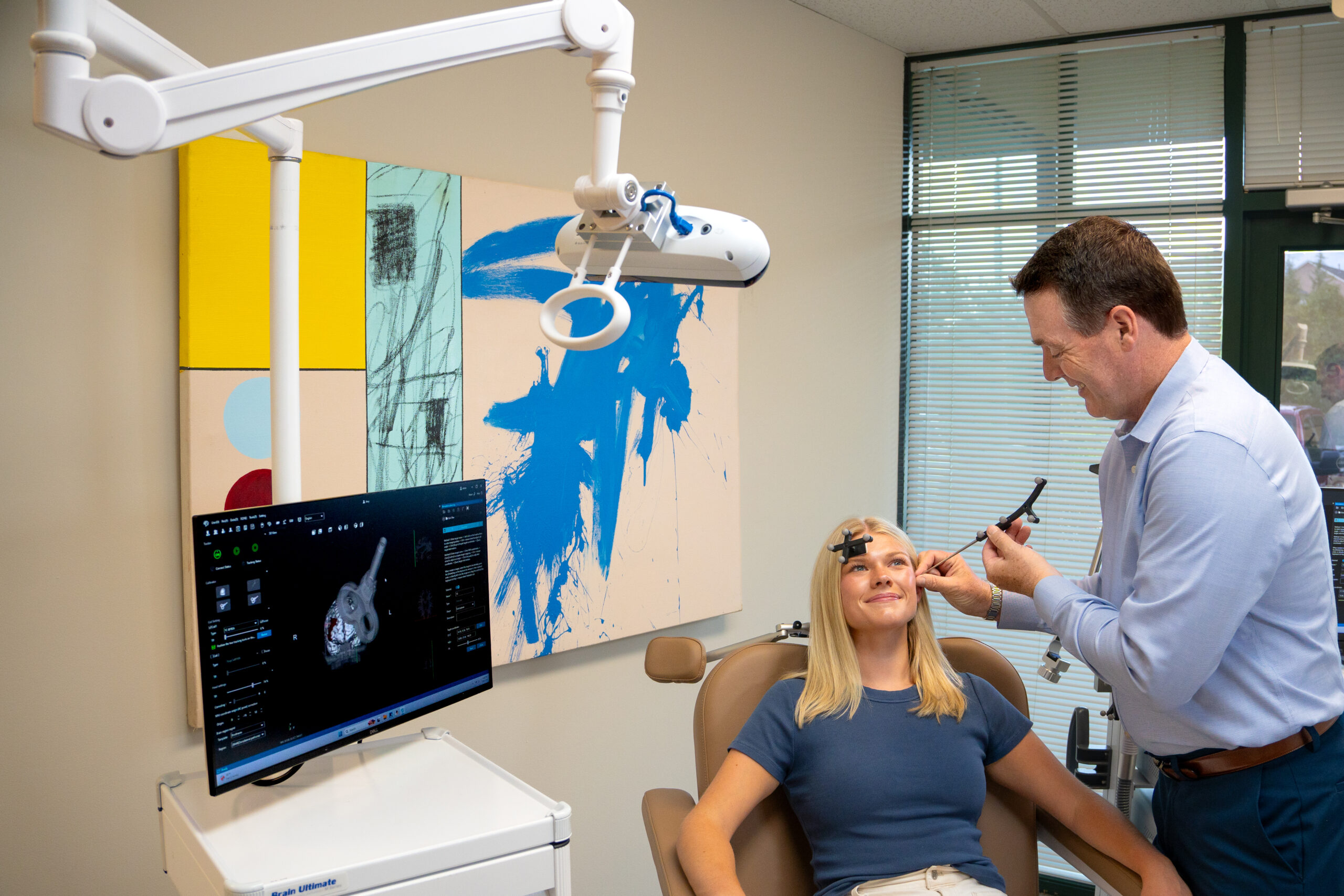
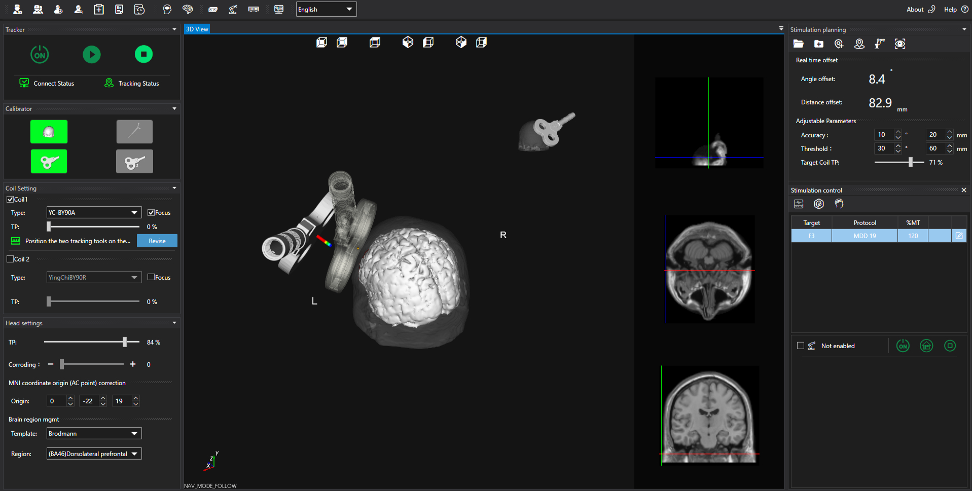
Brain Ultimate Neuronavigation uses an infrared optical tracking camera, mounted on an articulating arm for easy placement in any treatment room. This camera follows two small tracking tools in real time: one on the treatment coil, one on the patient's head. Both are calibrated to a 3D model of the patient's anatomy, so the system can show exactly where the coil sits relative to the intended target throughout the session.
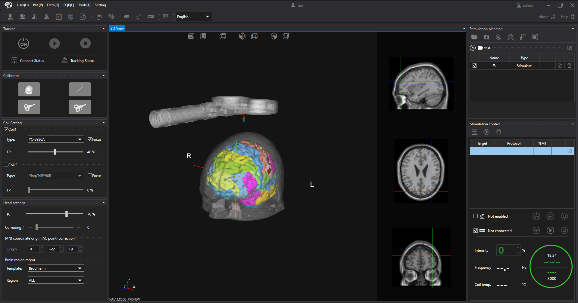
Everything runs through QuickVision, the software we built in-house to run navigation, targeting, stimulator control, and motor threshold determination all in one application. There's no second program to open and no separate record to update after the session.
A treatment target isn't just a location. Position coordinates tell you where on the scalp the coil sits, but orientation (pitch, roll, and yaw) determines how the coil is tilted and which way the magnetic field travels. Neuronavigation tracks all six values together, which is what makes a target reproducible from one session to the next. Our software will automatically adjust the target to the most effective coil orientation.
The system also supports loading a patient's own structural and functional imaging, in both DICOM and NIfTI formats. Either format is used to derive personalized MNI coordinates directly in the software. For patients without imaging, the system can use a common mode using standard adult male, adult female, adolescent, and child head models.
Five different ways to set a target are supported, from standard 10-20 sites and published MNI coordinates to the patient's own imaging, direct placement on the 3D model, or saving the coil's current position once calibrated.
Why It Matters
Head shape, cortical folding, and scalp-to-target depth all vary from person to person, so the same measurement can land in a meaningfully different place depending on the patient.
Neuronavigation replaces that estimate with a measurement. A target is chosen once, in the patient's own anatomy, and found again at every subsequent session rather than estimated each time. In practice, that gives clinics:
Live Guidance
Live guidance during positioning, with distance and angle displayed against a threshold zone.
Automatic Pause
Automatic pause if the coil drifts outside the threshold zone, resuming once it's repositioned.
Built-In Documentation
Documentation built into the workflow, with treatment records and reports generated.
Consistency
Consistency across staff and across a full treatment course, since a saved target and calibration carry over to a patient's next visit.
How It Supports Research
Reproducibility of coil placement is one of the main challenges in TMS research. Because Neuronavigation ties targeting to the patient's own imaging and tracks both position and orientation continuously, it gives research teams a way to:
- Derive personalized MNI coordinates from a subject's own structural or functional scan rather than approximating from a generic head shape
- Standardize targeting across visits, sessions, and operators, reducing a major source of variability in longitudinal or multi site protocols
- Produce a documented, session-by-session record of where and how each stimulation was actually delivered
Navigation supports the delivery of cleared Major Depressive Disorder protocols. Use outside those indications is for research only.
Availability
Brain Ultimate Neuronavigation is available now as an add-on for existing M-Series Ultimate systems or as part of a new system purchase. If you're interested in adding it to your clinic or research site, reach out to your Brain Ultimate representative to learn more about pricing, compatibility, installation, and training.
Contact Us